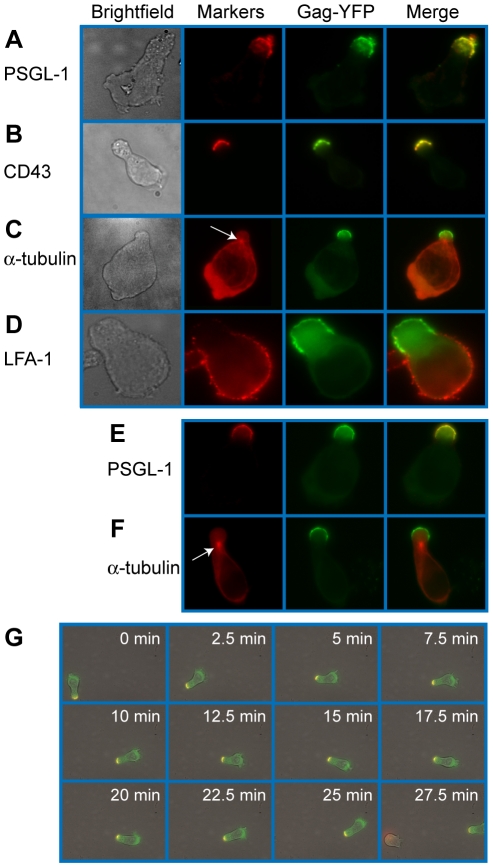
Figure 1. Gag stably localizes to the uropod in polarized T cells.
Primary T cells (A–D) and P2 cells (E–F) expressing Gag-YFP (green) were immunostained for uropod and non-uropod markers as described in the Materials and Methods section, and observed using an epifluorescence microscope. Uropods were identified by the presence of PSGL-1 (A and E) and CD43 (B) as well as by the location of the MTOC determined by immunostaining with anti- α-tubulin (C and F, arrows). LFA-1 (D) is a known non-uropod marker and served as a negative control. G) Cells expressing Gag-YFP (green) were immunostained with anti-PSGL-1 prelabeled by AlexaFluor-594-conjugated anti-mouse IgG (red). Images were acquired every 30 s for 30 min as the polarized cell migrated through the field. Yellow color indicates colocalization of PSGL-1 and Gag-YFP.

