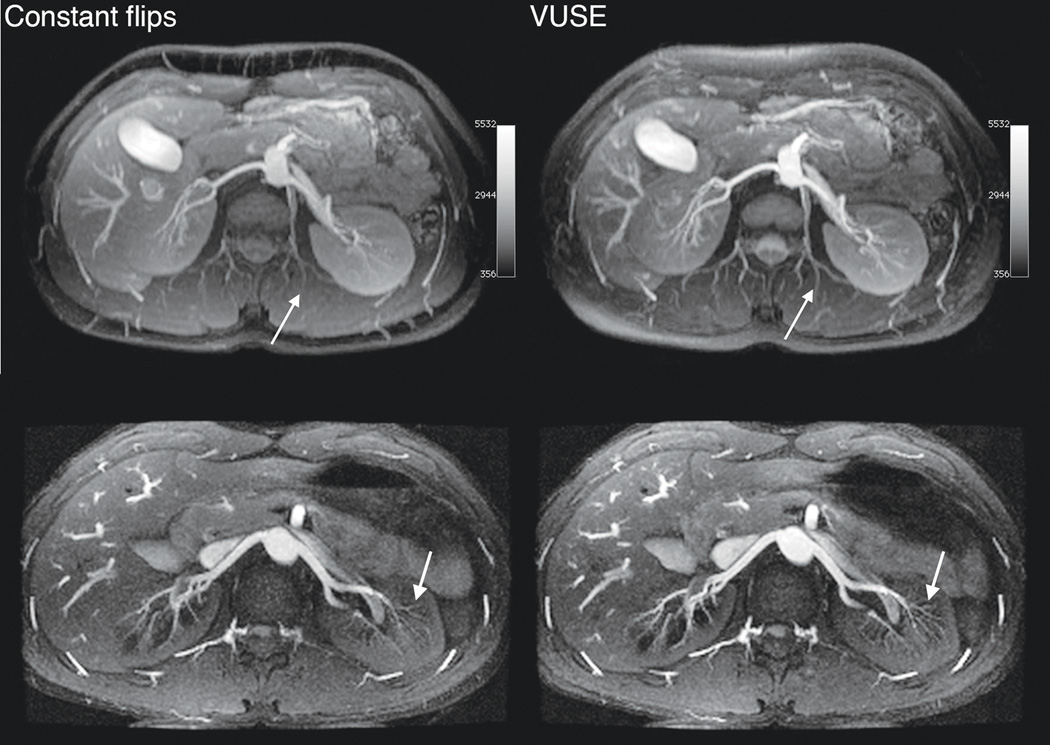
Figure 7.
Axial MIP renal angiograms from MRA acquisitions using constant flip angle (60°, upper left, and 70°, lower left) and VUSE bSSFP. In the second (bottom) volunteer, each kz plane was acquired in two acquisition blocks (ky interleaved). Images acquired using VUSE bSSFP has higher signal and improved small vessel depiction (white arrows). Each row is displayed at the same window and level.

