
Figure 1.
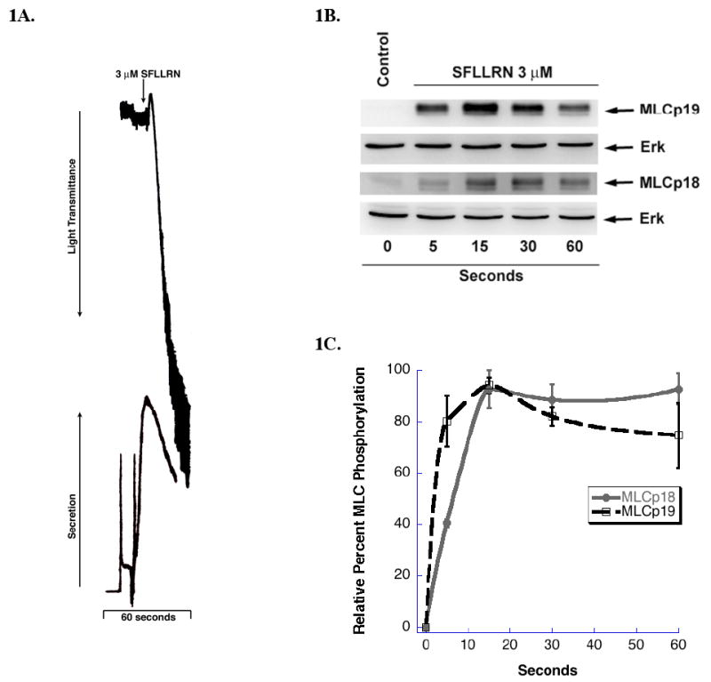
Phosphorylation of MLC by PAR 1 agonist (A) Washed and aspirin-treated human platelets where stimulated with 3 μM SFLLRN at 37° C while stirring for 60 seconds. Platelet aggregation and secretion where measured in a Lumi-aggregometer. The tracings are representative of data from at least three independent experiments. (B) SFLLRN stimulated platelets were subjected to 15% SDS-PAGE and analyzed for MLC phosphorylation by using polyclonal anti-phospho-specific (Ser) 19 or anti-phospho-specific (Thr)18 antibodies. Equal lane loading was assured by probing the samples with total monoclonal Erk antibody. (C) Densitometrical analysis of the western blot was performed. Each data point is the mean ± S.E. percentage value (n = 3) of phosphorylated MLC. The samples that make up each of these data points are derived from platelets from 3 different donors.