Fig. 3.
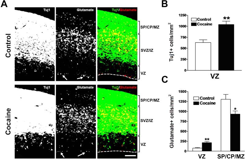
Effects of cocaine on the densities of Tuj1- and glutamate-positive cells in the neocortex during the middle period of neocortical neurogenesis. (A) Immunostaining for Tuj1 (green) and glutamate (red) in the neocortex of control and cocaine-exposed fetuses. Glutamate-like staining at the ventricular surface, which is often seen at the edges of the sections, was excluded from quantification (white arrows show examples). Scale bar = 100 μm. VZ, ventricular zone; SVZ, subventricular zone; IZ, intermediate zone; SP, subplate; CP, cortical plate; MZ, marginal zone. (B) Tuj1-positive cells in the VZ were increased in cocaine-exposed fetuses. **p<0.01 compared to control, n = 8 per group from three individual pregnant dams. (C) Cocaine exposure caused an increase in the density of glutamate-positive cells in the neocortical VZ, but a decrease in the SP/CP/MZ. *p<0.05, **p<0.01 compared to control, n = 8 per group from three individual pregnant dams.

