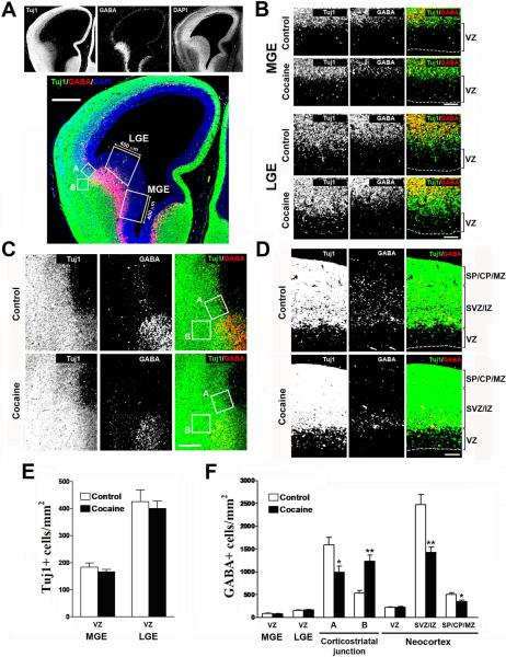
Fig. 4.
Effects of cocaine on the density of GABA-positive cells in the fetal cortex during the middle period of neocortical neurogenesis. (A) Immunostaining for Tuj1 (green), GABA (red), and DAPI (blue) in the fetal cortex. The boxed areas (400 μm in width) mark the medial and lateral ganglionic eminences (MGE and LGE), and were used for quantification of the densities of Tuj1- and GABA-positive cells. The square boxes (A and B, 150×150 μm) were used for quantification of the GABA-positive cells either migrating tangentially to the neocortex (Box A) or accumulating in the ventral telencephalon below the corticostriatal junction (Box B). Scale bar = 400 μm. (B) Immunostaining of Tuj1 (green) and GABA (red) in the MGE and LGE of control and cocaine-exposed fetuses. Scale bar = 100 μm. VZ, ventricular zone. (C) Immunostaining of Tuj1 (green) and GABA (red) in the corticostriatal junction of control and cocaine-exposed fetuses. Scale bar = 200 μm. (D) Immunostaining of Tuj1 (green) and GABA (red) in the neocortex of control and cocaine-exposed fetuses. GABA-like staining at the ventricular surface that is often seen at the edge of the section was excluded from quantification (white arrows show examples). Scale bar = 100 μm. VZ, ventricular zone; SVZ, subventricular zone; IZ, intermediate zone; SP, subplate; CP, cortical plate; MZ, marginal zone. (E) Densities of Tuj1-positive cells in the VZ of the MGE and LGE were unaltered by cocaine exposure. N = 8 per group from three individual pregnant dams. (F) Densities of GABA-positive cells were unchanged in the VZ of the MGE and LGE in cocaine-exposed fetuses. Cocaine exposure caused the densities of GABA-positive cells to decrease in the neocortical SVZ / lower IZ (Box A), but concomitantly increase in the subcortical region (Box B). Quantification of GABA-positive cells showed no differences in the neocortical VZ as compared to control, but a decrease in both the SVZ/IZ and SP/CP/MZ. *p<0.05, **p<0.01 compared to control, n=8 per group from three individual pregnant dams.

