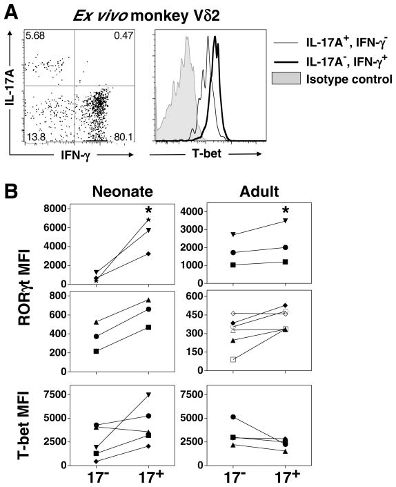
FIGURE 6. Expression of RORγt and T-bet by IL-17A+ Vγ2Vδ2 T cells.
A. Representative staining for T-bet on monkey peripheral blood Vδ2 T cells, segregated into IL-17A+, IFN-γ− Vδ2 T cells (Tγδ17) or IL-17A−, IFN-γ+ Vδ2 T cells (Tγδ1). Represents one of three monkeys examined. PBMC were isolated and stimulated with PMA and ionomycin and intracellular staining for IL-17A, IFN-γ, and T-bet performed. B. (Neonate, left) Cord blood mononuclear cells were polarized with HMBPP for 13 days in the presence of IL-1β, IL-6, TGF-β, and anti-IL-23, and (Adult, right) adult PBMC were polarized with HMBPP for 7 days in the presence of IL-1β, IL-23, TGF-β, and anti-IL-6. On the final day, cells were restimulated with PMA and ionomycin, surface stained for Vδ2 and CD3, and intracellularly stained for IL-17A, RORγt, and T-bet. Vγ2Vδ2 T cells were segregated into IL-17A+ and IL-17A− and the MFI for each transcription factor minus the MFI of the respective isotype control is shown. Because donors had variable baseline RORγt staining, the donors are segregated into two graphs to accommodate the different magnitudes exhibited. Note that cells were not segregated based on IFN-γ production, therefore the neonatal IL-17A+ fraction refers to the sum of Tγδ1/17 and Tγδ17. * p < 0.05, Kruskal Wallis comparison with IL-17A− group.

