
Figure 3. Neural progenitor cells in WT and Nova2 KO cortex.
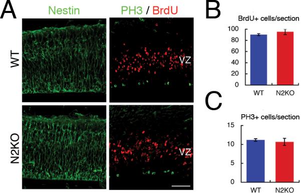
(A) BrdU was injected 1 hr before fixation. Coronal sections of embryonic day 14.5 cortex in wild type and Nova2 KO brains were immunostained with antibodies against Nestin (left panel; green), which is neural progenitor marker protein, BrdU (right panel; red) and PH3 (right panel; green). (B) This graph represents the number of BrdU positive cells per section. (C) This graph represents the number of PH3 positive cells per sections. Error bar indicates SD of three biological replicates. Scale bar; 50 μm. PH3; phospho-Histone3.