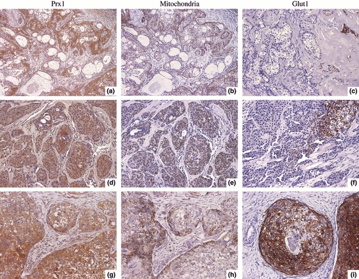
Figure 1.

Immunohistochemical expression of Prx I, mitochondrial antigen and glucose transporter protein 1 (Glut-1) in Mucoepidermoid carcinoma (MEC) presenting different histological grades. (a–c) Low-grade MEC. (a, b) Positive Prx I and mitochondrial antigen staining. (c) No reactivity to Glut-1, except in the scanty tumour cells scattered within hyalinized area (arrow). (d–f) Intermediate grade MEC. (d, e) Malignant cell blocks exhibit reactivity to Prx I and mitochondria antigen. (f) Membranous Glut-1 expression in a focal area. (g–i) High-grade MEC. (g, h) Positive Prx I and mitochondrial antigen staining. (i) Malignant cell blocks frequently exhibit intense membranous Glut-1 reactivity. (Original magnifications ×400, except a, b ×200).
