
Figure 6.
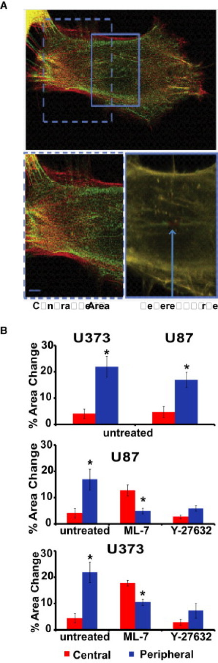
Effect of pharmacological inhibition of MLCK and ROCK on cell area over long timescales. (A) Effect of MLCK inhibition on central fibers. In cells treated with 5 μM of ML-7, photodisruption of a central fiber induces a dramatic change in area, whereas photoablation of a remaining peripheral fiber shows a negligible change in area. All images are overlays of mCherry-LifeAct fluorescence before ablation (red) and 30 min postablation (green). The bottom images are high-magnification zooms of the boxed regions in the top image. (B) Quantification of area changes as a function of location of the severed fiber. Top panel: Area changes for untreated control cells. Middle panel: Area changes in U87 cells treated with ML-7 and Y27632. Bottom panel: Area changes in U373 cells treated with ML-7 and Y27632, emphasizing the transference of tension in response to ablation of SF subpopulations. ∗ Statistically significant area changes (p < 0.01) as determined by a Student's t-test (unpaired, two-tailed, 95% confidence interval). Error bars represent the mean ± SE for central versus peripheral for each condition. N > 20 SFs per condition were ablated.