
Figure 2.
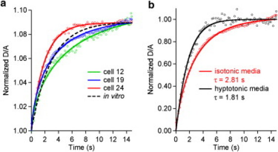
(a) Protein folding/unfolding relaxation monitored near the denaturation midpoint (jump starting at 39°C) in three of the 30 studied cells compared to aqueous solution (dotted black line). The parameter obtained from fits to stretched exponentials are summarized in the Supporting Material. (b) Cells with greater water content (hypotonic) promote faster protein folding.