
Figure 2.
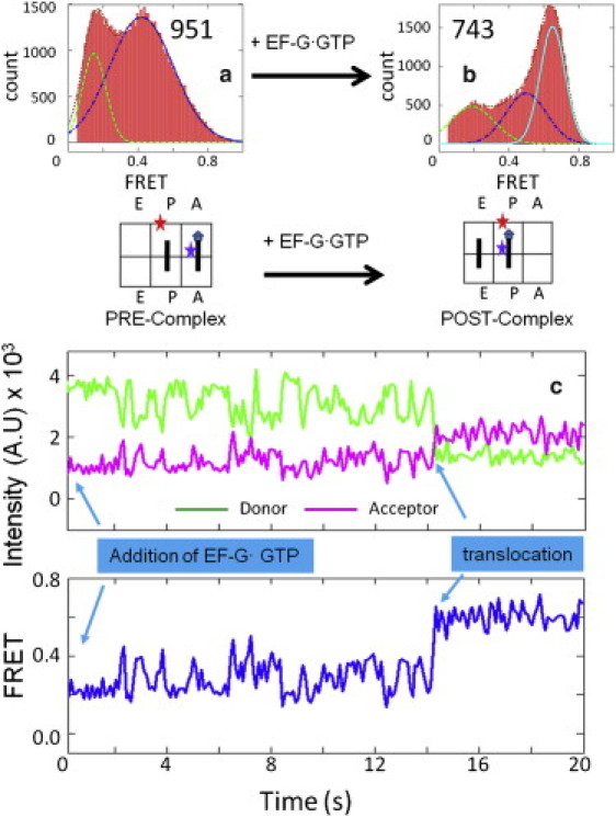
FRET distributions of the signals between the L27 protein and the A-/P-site tRNAs in the (a) Pre-complex (951 particles) and (b) Post-complex (743 particles). The sketches beneath the histogram plots show the relative position of the FRET pairs. (c) One representative trace of the real-time observation of the translocation process. Imaging began immediately after EF-G·GTP (200 nM) was loaded onto the surface-bound Pre-complex. Translocation happened after ∼14 s.