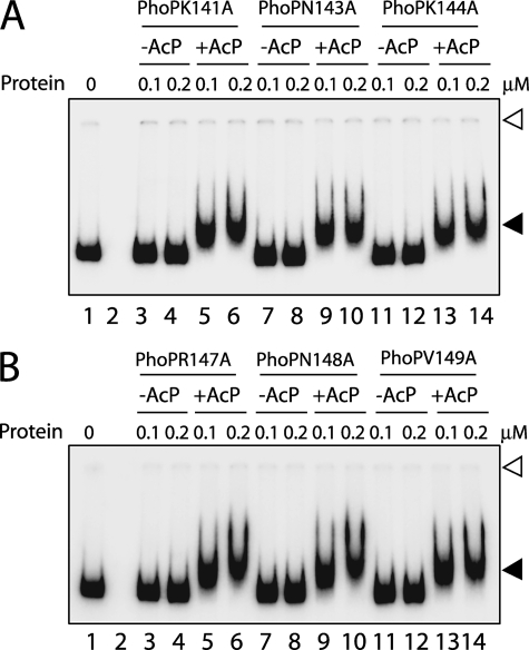
FIGURE 6.
A and B, EMSA of radiolabeled msl3 promoter region for binding of the indicated PhoP linker mutants carrying single alanine substitutions. Sample analysis and detection of protein-DNA complexes were as described in the legend to Fig. 1. Open and filled arrows indicate origins of the polyacrylamide gel and retarded complexes with band shifts produced in the presence of PhoP proteins, respectively. The gels are representative of at least three independent experiments.

