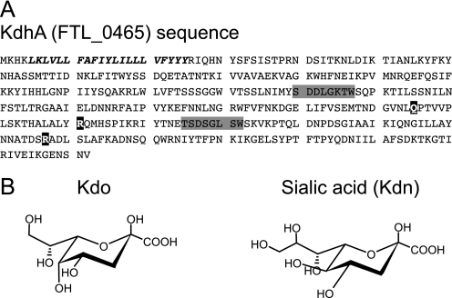
FIGURE 1.
A sialidase-like protein in F. tularensis LVS. A, amino acid sequence of F. tularensis LVS KdhA showing the transmembrane domain (italics), the BNR/Asp repeats ((S/T)XDXGXT(W/F)) of bacterial sialidase (shaded in gray), and three of the seven residues that compose sialidase catalytic site (in black). Domain predictions were performed using CD-Search (45). B, chemical structure of Kdo and sialic acid Kdn.

