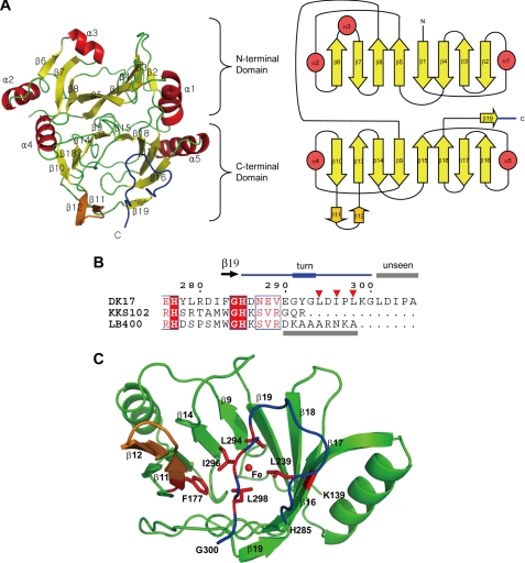
FIGURE 2.
Monomeric structure of AkbC. A, ribbon (left) and topology (right) diagrams of the AkbC monomer. B, sequence alignment of the C-terminal tail. Residues Arg-276—Ala-305 form AkbC is aligned with those of DHBDs from Pseudomonas sp. strain KKS102 (Arg-276—Arg-292) and B. cepacia LB400 (Arg-276—Ala-298). C, C-domain structure of AkbC. Hydrophobic residues in the β-hairpin structure (β11–β12, orange) and the C-terminal tail (blue) are shown as red sticks and marked as inverted triangles. Unseen regions in the crystal structures are indicated with gray bars.

