FIGURE 7.
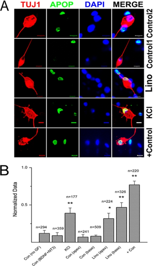
Qualitative assessment of SGNs apoptosis. A, upper panel shows SGNs in control(2) culture with BDNF/NT3 growth factors. Note that there are non-neuronal cells undergoing apoptosis, but invariably, most of the SGNs were intact. The 2nd upper panel is from control(1) culture with no growth factors (GF). The number of apoptotic SGNs increased when cultures were treated with 10 μm linopirdine (lino, 3rd row) and 10 mm KCl for 72 h. The bottom row serves as a positive control (Con) with treatment of 1 μm 12-dimethylbenz[a]anthracene. TUJ1, neuronal marker; Apop (apoptosis = TUNEL-positive; DAPI = nuclei stain). B, quantitative assessment of SGNs apoptosis histograms, comparing the ratio of TUNEL-positive SGNs under different experimental conditions. Control 1, have no growth factor; control 2, contained BDNF/NT3, 10 mm, KCl, 10 μm linopirdine (lino), comparison of base-line data for apical and basal SGNs with linopirdine-treated conditions, and finally, after application of positive control = 1 μm 12-dimethylbenz[a]anthracene. **, p < 0.01.

