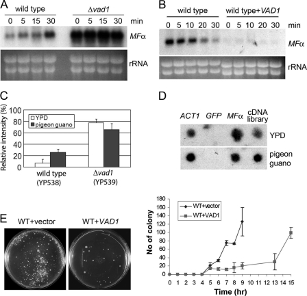
FIGURE 5.
Accumulation of both MFα1 transcript and MFα1-GFP fusion protein on pigeon guano medium. A, Northern blot analysis of steady state levels of MFα1 of wild type and Δvad1 mutant cells upon transition to pigeon guano medium. RNA was prepared at the indicated times after transfer of cells from YPD to pigeon guano medium. Membranes were hybridized with MFα ORF sequences. B, wild type or VAD1-overexpressing strains (mid-exponential phase) were transferred to pigeon guano medium, and mRNA degradation was assayed. C, relative intensity of MFα1-GFP cultured on YPD or pigeon guano medium, measured by fluorescence microscopy as in Fig. 3A. D, transcriptional run-on analysis of MFα1 in YPD and after a 30-min incubation on pigeon guano medium. Dot blots were prepared with PCR-amplified fragments (10 μg/sample) using primers from within the coding regions of the respective gene targets. E, fusion mating assays were performed as described under “Experimental Procedures.” Agar plates were photographed at the indicated time points (left, 13 h), and fused colony numbers of indicated strains were counted and recorded (right). Error bars, S.E.

