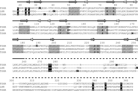
FIGURE 1.
Alignment of the three human GpHR ECDs. The numbering given above the sequence is based on FSHR. The arrows, positioned on the solid line (above the numbering) that represents the part of the FSHR of which the structure is known (30), indicate the β-strands on the concave (dark gray) or convex (light gray) side of the FSHR ECD. The dashed line (above the numbering) indicates the part of the FSHR ECD of which no structural information is known yet. The invariant and highly conserved amino acid residues that were studied are shown in boldface, and all cysteine residues are presented in white on a darker background. Residues on a gray background are forming the β-strands facing the hormone in FSHR and are presumed to do the same in LHR and TSHR.

