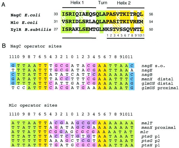
Figure 1.
Comparison of NagC and Mlc DNA binding motifs and operators. (A) Alignment of the HTH motifs in the N-terminal regions of NagC and Mlc from E.coli and the XylR repressor from Bacillus subtilis. The locations of the motifs relative to the N-terminal of the proteins are given. Amino acids in the recognition helix (helix 2 shown in yellow) are numbered 1–11. Identical amino acids are indicated by shading and conservative replacements by hatching. (B) Alignment of native NagC and Mlc operator sites. The conserved TT/AA bases at positions –5,6/+5,6 are shown in yellow. AT bases in positions 7–10 (NagC) or 7–11 (Mlc) are shown in green, while C or G at positions +11/–11 of NagC are in blue. C or G residues in pink are in positions –4 to +4, which conforms to the CGCGNCGCG pattern, while those in mauve are in the GCGCNGCGC order.

