FIGURE 6.
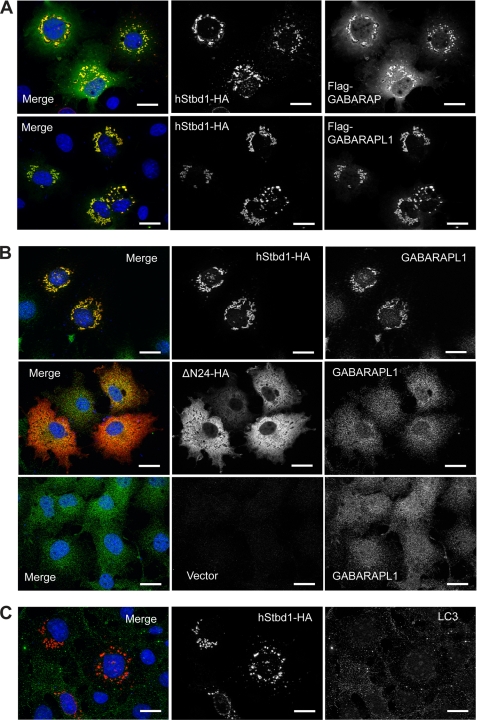
Subcellular localization of GABARAPL1, GABARAP and LC3 in relation to Stbd1. A, C-terminal HA-tagged hStbd1 was co-expressed in COS M9 cells with N-terminal FLAG-tagged GABARAPL1 or GABARAP, as in Fig. 5, and immunostained with anti-HA antibodies (red) or anti-FLAG antibodies (green). B, HA-tagged Stbd1 or N-terminally truncated Stbd1 was expressed in COS M9 cells and immunostained with anti-HA antibodies to detect Stbd1 (red) and anti-GABARAPL1 antibodies to visualize endogenous GABARAPL1 (green). The bottom row shows cells transfected with empty vector (pcDNA3) to reveal the endogenous GABARAPL1 distribution (green). C, HA-tagged Stbd1 was expressed in COS M9 cells and immunostained with anti-HA antibodies (red) or anti-LC3 antibodies (green) to detect endogenous LC3. Nuclei were stained with Hoechst (blue). Scale bar, 20 μm.

