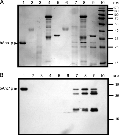
FIGURE 2.
Proteolysis of bAnc1p. Pepsin, type XIII, and type XVIII proteases (commercial and recombinant) were tested, and the corresponding lysates were analyzed by SDS-PAGE (12.5% acrylamide) and revealed by Coomassie Blue staining (A) or Western blot with anti-SDS-bAnc1p (B). Lanes 1–5, 2 μg of bAnc1p, 2 μg of pepsin, 26 μg of type XIII protease, 32 μg of commercial type XVIII protease, and 2 μg of activated recombinant type XVIII protease were incubated in acidic buffer for 2 min at 4 °C. Lanes 6–9, digestion assays were performed with 2 μg of bAnc1p and protease/substrate ratio of 1:1 (w/w) for pepsin, 10:1 (w/w) for type XIII protease, 17:1 (w/w) for commercial type XVIII protease, and 1:1 (w/w) for recombinant type XVIII protease in acidic buffer during 2 min at 4 °C, respectively. Lane 10, prestained molecular weight markers. The position of bAnc1p is indicated by an arrowhead.

