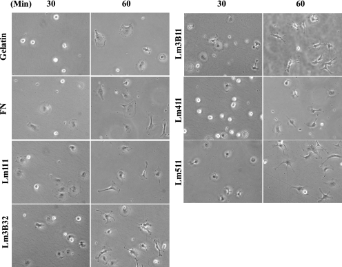
FIGURE 7.
Morphological change of MVE cells on Lm3B11 and other cell adhesion substrates. MVE cells (1 × 10⁴ cells in MCDB131 plus 1% FCS) were plated onto 24-well culture plates precoated with 40 μg/ml gelatin, 10 μg/ml fibronectin (FN), 10 μg/ml Lm111, 1 μg/ml Lm3B32, 5 μg/ml Lm3B11, 5 μg/ml Lm411, or 2.5 μg/ml Lm511 and incubated. Phase-contrast micrographs were taken 30 and 60 min after plating. Original magnification, ×200.

