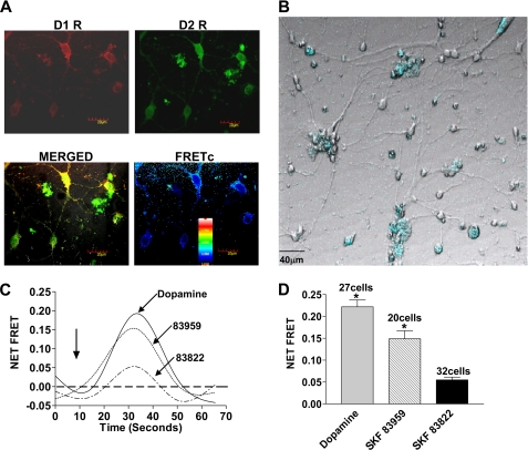
FIGURE 4.
Specificity of dopamine receptor agonists activating the D1-D2 receptor heteromer calcium signal in primary striatal neurons. A, immunocytochemistry shows endogenously expressed dopamine D1 and D2 receptor colocalization (merged) and interaction (corrected FRET (FRETc)). The inset shows the calibration for FRET efficiency. B, striatal neurons display the presence of transfected cameleon (blue). C, representative tracings of cameleon FRET result from intracellular calcium release from D1-D2 receptor heteromer activation by 100 nm dopamine or SKF 83959 but not SKF 83822. The arrow indicates the time point of drug addition. D, peak heights of agonist-induced cameleon FRET correspond to calcium release through activation of the D1-D2 receptor heteromer. The results shown represent the means ± S.E. of values from the number of cells shown. A significant difference from SKF 83822 is denoted by * = p < 0.05.

