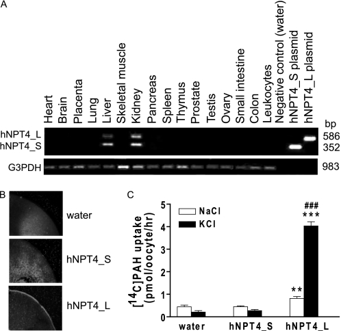
FIGURE 2.
Tissue distributions of human NPT4 and its function. A, distributions of NPT4 mRNA in human multiple cDNA panels. mRNA expression of two isoforms for hNPT4 were detected in the kidney and liver. Control amplification with GAPDH was performed in parallel (bottom panel, G3PDH). B, subcellular localization of hNPT4 short (hNPT4_S) and long (hNPT4_L) isoforms in oocytes. Immunodetection with a specific antibody, raised against the N terminus of hNPT4 (Sigma), showed that hNPT4_L is expressed at the plasma membrane, whereas fluorescence levels were undetectable on plasma membrane of oocytes injected with water or hNPT4_S cRNA. C, [14C]PAH uptake by oocytes injected with water, hNPT4_S, and hNPT4_L. The oocytes were incubated in basal uptake solution (96 mm NaCl, 2 mm KCl, 1.8 mm CaCl2, 1 mm MgCl2, 5 mm HEPES, pH 7.4) (open columns) and high potassium uptake solution (Na+ in the basal uptake solution was replaced by K+) (closed columns) containing 10 μm [14C]PAH for 1.0 h. The data are the means ± S.E. with n = 6–8. **, p < 0.01; ***, p < 0.001 versus the uptake of water-injected oocytes; ###, p < 0.001 versus the uptake in basal (NaCl) solution.

