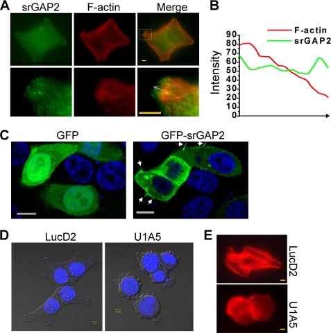
FIGURE 2.
srGAP2 localizes to sites of membrane protrusion and is required for cell morphology. A, HCT116 cells at 24 h after seeding on the coverslip were double labeled with mouse anti-srGAP2 antibody, followed by anti-mouse FITC-conjugated secondary antibody and TRITC 545-phalloidin. Bars, 10 μm. The lower figures are the high-magnification from the upper selected inset, showing that srGAP2 localized to leading edge of F-actin at the cell membrane protrusion. B, graphs correspond to intensities in arbitrary units of the green (srGAP2) and red (F-actin) labeling for each pixel of the arrow drawn through the axis in A. C, HeLa cells transfected with GFP or GFP-srGAP2. The arrow indicated filopodia-like protrusion. Bars, 10 μm. D and E, srGAP2 regulates the cell morphology. Graphs taken at 33 h after seeding, LucD2 (control) showed well-pronounced protrusions and spread well, U1A5 (srGAP2 depleted) became rounded. DNA was stained with DAPI (blue) and visualized by DIC (D). Cells were stained with TRITC 545-phalloidin to visualize F-actin (red) (E). Bars, 10 μm.

