
Fig. 2.
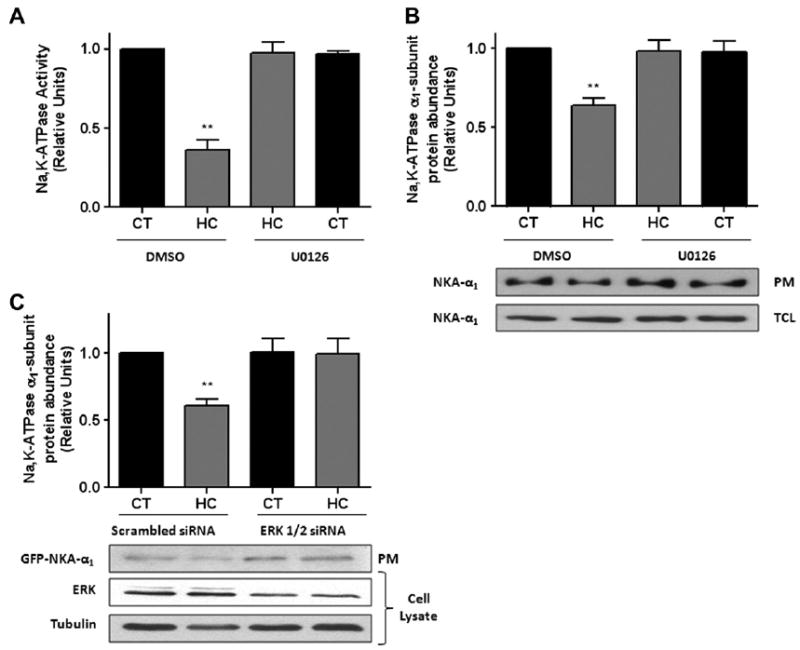
Inhibition of ERK 1/2 prevents the hypercapnia-induced Na,K-ATPase downregulation. (A) ATII cells were pre-treated with U0126 (10 μM) or DMSO for 30 min and then exposed to 40 (black bars) or 120 (grey bars) mmHg pCO2 for 30 min in the presence or absence of U0126. Na,K-ATPase activity was measured as [γ-32P] ATP hydrolysis. (B) ATII cells treated as in (A) and Na,K-ATPase protein abundance at the plasma membrane (PM) was determined by biotin-streptavidin pull down and subsequent Western blot against Na,K-ATPase α1-subunit. Total cell lysates are also shown. (C) A549-GFP-α1 cells were transiently transfected with scrambled or ERK 1/2 siRNA and 48 h later exposed to 40 or 120 mmHg pCO2 for 30 min. The Na,K-ATPase protein abundance at the plasma membrane (PM) was determined as in (B). Representative Western blots of total cell lysates for total ERK 1/2 and tubulin are shown. Graph represents the mean ± S.E.M, n = 5. **P < 0.01.