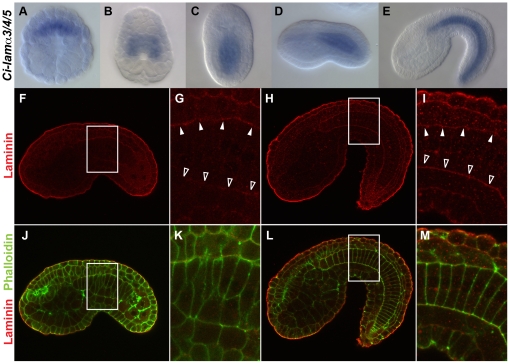
Figure 1. Distribution of laminin in Ciona intestinalis embryos.
(A–E) Whole-mount in situ hybridization of early gastrula (A), late gastrula (B), late neurula (C), early tailbud (D) and middle-late tailbud (E) stages for Ci-lamα3/4/5 mRNA. A is vegetal view, B and C are dorsal views, and D and E are lateral views. Anterior is to the top in A–C and to the left in D and E. Note that Ci-lamα3/4/5 is preferentially expressed in notochord lineage cells. (F–M) Confocal images of embryos stained with the anti-Cs-lamα3/4/5 (red) alone (F–I) and double staining with phalloidin (green, J–M) at early tailbud (F,G,J,K) and middle-late tailbud (H,I,L,M) stages. All images are lateral views and anterior is to the left. G, I, K and M are higher magnification of the boxed area in F, H, J and L, respectively. Note: In all cases, high background red signal outlining embryos was seen perhaps due to an edge effect as Cs-lamα3/4/5 mRNA is undetectable there. At early taulbud stage, a high signal accumulation or maternal laminin protein is detectable at the dorsal side (white arrowheads in G) but not at the ventral side (open arrowheads in G) of the notochord. When intercalation is completed, Cs-lamα3/4/5 signal expands to the ventral side (open arrowheads in I) of the notochord and asymmetric distribution along the dorsoventral axis is lost (I,M).

