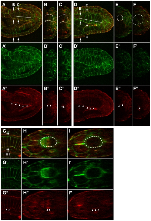
Figure 3. Accumulation of aPKC at the ventral surface of notochord cells is retained during cell intercalation process.
(A,D,G) Confocal saggital section images of the notochord of a late neurula (A), early tailbud (D) and middle-late tailbud (G) stage embryos double-stained for aPKC (red) and phalloidin for actin (green). Lateral views with anterior to the left. Reconstructed cross section image at the level indicated by arrows in A, D and G is shown in B/C, E/F and H/I. Position of the notochord in saggital section and cross section images is indicated by a white line in A and D or surrounded by white dotted line in B,C,E,F,H,I. Accumulation of aPKC is indicated by white arrowheads in A’–I’.

