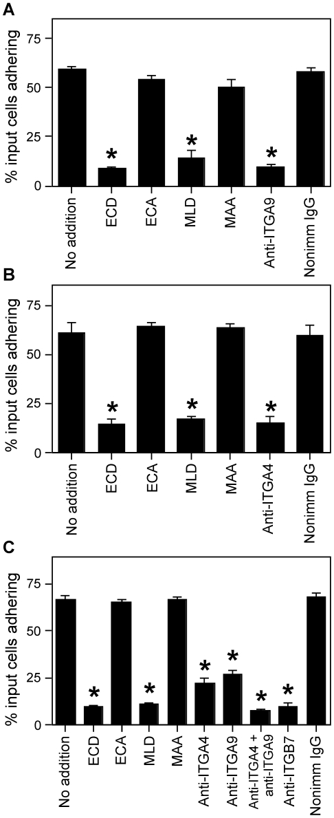
Figure 3. ITGA4/ITGA9-mediated cell adhesion to ADAM2.
Cell adhesion assays were performed with the indicated cell lines (Panel A, Tera-2 cells; Panel B, HT1080 cells; Panel C, RPMI 8866 cells) with 50 µg/ml of recombinant ADAM2 as substrate. Cells were left untreated (no addition), or treated with the indicated peptide (100 µM; ECD peptide, corresponding to the ADAM2 disintegrin loop; its negative control ECA; the ITGA4/ITGA9-blocking peptide MLD, or its control MAA) or the indicated antibody (20 µg/ml; function-blocking anti-ITGA9 monoclonal antibody Y9A2 [Panels A and C]; the function-blocking anti-ITGA4 monoclonal antibody PS-2 [Panels B and C]; function-blocking anti-ITGB7 monoclonal antibody FIB27 [Panel C]; or a species-matched nonimmune IgG). The y-axes indicate the percentage of the input cells left adherent after washing; errors bars represent the SEM. Asterisks indicate p<0.05 as compared to untreated controls and the appropriate control peptide or nonimmune antibody.

