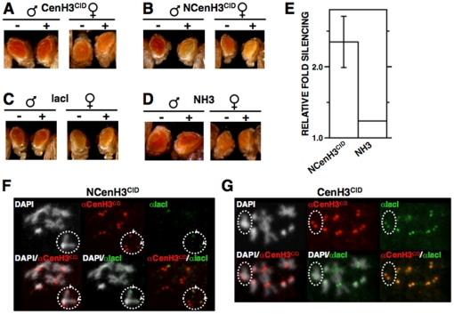
Figure 1. Tethering NCenH3CID to a white-reporter silences reporter expression.
(A–D) The eye phenotype of S9.2 flies expressing the indicated lacI-fused proteins (+) is compared to that of siblings where no fused protein is expressed (−). Results are presented for both female and male individuals. (E) Quantitative analysis is presented for lines expressing NCenH3CID-lacI and NH3CID-lacI constructs. Relative fold silencing is expressed as the ratio between OD480 of control S9.2 lines expressing no fused protein and that of lines expressing the indicated constructs. For NCenH3CID-lacI, results correspond to the average of three independent lines. For NH3CID-lacI, results are presented for a single representative line. (F and G) NCenH3CID-lacI, but not CenH3CID-lacI, bind to the ectopic white-reporter construct. Fused proteins were expressed in 157.1 flies, where the white-reporter is inserted at a distal position on the X-chromosome, and localisation was determined in mitotic chromosomes by immunostaining with αlacI (green) and αCenH3CID (red), which also detects endogenous CenH3CID at centromeres. Dotted circles indicate X-chromosomes. Arrows indicate co-localisation of αCenH3CID and αlacI signals at ectopic sites on the X-chromosome, reflecting binding of NCenH3CID-lacI to the reporter. DNA was stained with DAPI. See Figure S4 for a description of the constructs.

