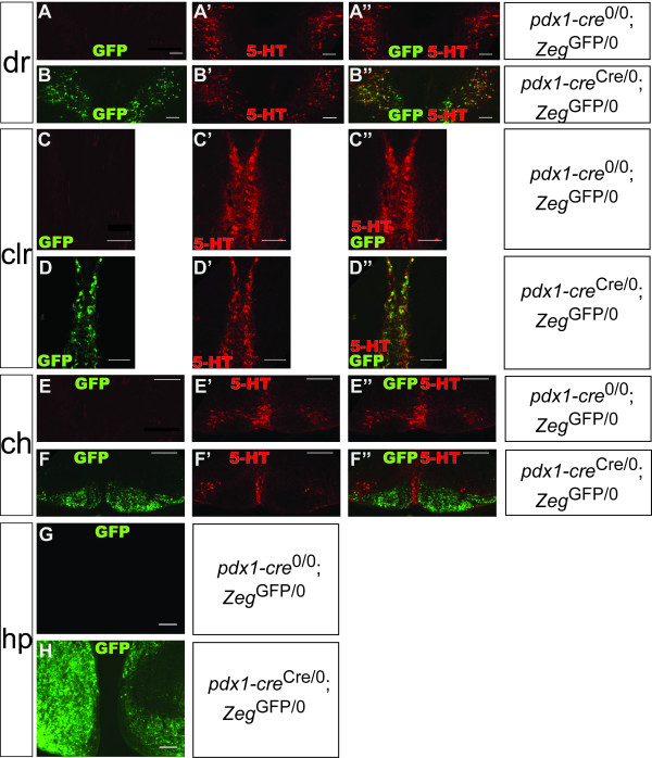
Figure 4.
Cre-mediated recombination in the hindbrain and diencephalon in the e16.5 embryo. Epifluorescence images from pdx1-creCre/0; ZegGFP/0 (B, D, F, H) and pdx1-cre0/0; ZegGFP/0 (A, C, E, G) e16.5 embryos, transversely sectioned, immunostained for GFP (green) and 5-HT (red). GFP was expressed in the dorsal raphe nucleus (dr) (B), caudal linear raphe (clr) (D), caudal hindbrain (ch) (F) and hypothalamus (hp) (H). In the rostral hindbrain, GFP expression occurred in the serotonergic dorsal raphe and caudal linear nuclei (B, D). In the caudal hindbrain, GFP expression was observed in the non-serotonergic inferior olive nucleus, adjacent to serotonergic raphe nuclei (F). GFP expression was not evident in sections from pdx1-cre0/0; ZegGFP/0 control embryos (A, C, E, G). Scale bars, 100 μm.

