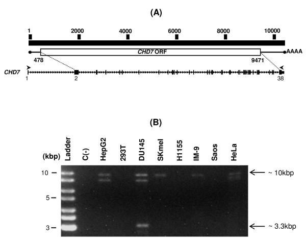
Figure 1.
Identification of a putative novel splice variant of CHD7. (A) Representation of the full-length CHD7 transcript structure and primers used to amplify it. At the top the nucleotide sequence numbering scheme of the full-length transcript is represented. At the middle is a schematic representation of the full-length CHD7 transcript with its ORF indicated as a box (the ORF initial and final nucleotides numbers are indicated below the box). At the bottom the exon-intron structure of CHD7 is represented. Exons are represented as black rectangles and intronic sequences as a thin line. The sites of the ORF initial and final nucleotides (dotted lines) and the primers (arrows) annealing to sequences corresponding to exons 1 and 38 of the CHD7 transcript sequence (deposited under the accession number NM_017780.2) used for the amplification of the full-length CHD7 transcript are indicated. (B) Amplification of the full-length CHD7 transcript by RT-PCR. Poly A+ RNA was extracted from the indicated cell lines, and reverse transcription followed by PCR was performed using the CHD7 primers set depicted in (A). Aside from the predicted 10 kbp band, additional RT-PCR products of approximately 9 kbp (not indicated) and 3.3 kbp (indicated) were detected. C(-) is the negative control (PCR reaction without cDNA).

