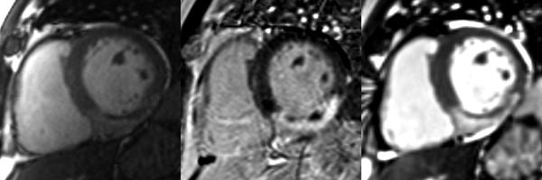
Figure 1.
Matched diastolic cardiac MRI (left - cine MRI; middle - phase sensitive inversion recovery image; right - T2-weighted steady state free precession [SSFP]) obtained in a 57 year-old man one week after primary PCI to the right coronary artery for an acute inferior STEMI. Post-PCI culprit artery flow was reduced (TIMI flow grade 1). Transmural infarction (as revealed by late gadolinium enhancement, middle) corresponds with transmural edema (bright blood T2-weighted SSFP, right). Absolute AAR and salvage in this patient were 29% and 6%, respectively. The central dark zones within the infarct territory represent MVO complicated by hemorrhage (28).

