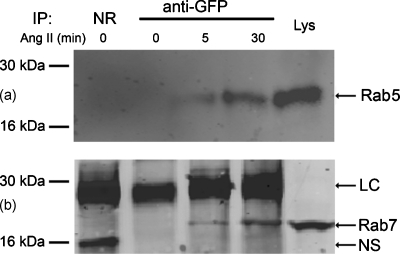
Figure 4.
Coimmunoprecipitation of AT1R-EGFP with (a) Rab5 and (b) Rab7 in AT1R-HEK293 cells. Cells were treated with Ang II (100 nM), at the indicated periods. One mg of cell lysates were incubated with anti-GFP IgG at 4 °C. The precipitated protein complexes were immunoblotted with (a) Rab5 mAb or (b) anti-Rab7 IgG. Both Rab5 and Rab7 coimmunoprecipitated with AT1R-GFP in the samples treated with Ang II for 5 min and 30 min. NR, normal rabbit IgG; LC, IgG light chain; NS, nonspecific band; Lys, whole cell lysate.

