
Figure 1. Permeabilized cell nuclear import assay adapted to 96-well plate format.
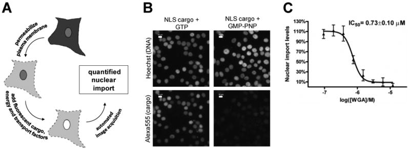
A. Schematic diagram of permeabilized cell nuclear import assay. Soluble cytosolic components are released after the plasma membrane is perforated by the glycoside digitonin, which leaves the NE and ER membranes intact. An ATP regenerating system, GTP, nuclear transport factors and labeled cargo is added exogenously. The fluorescence accumulated in the nucleus is quantified by light microscopy. B. Fluorescence microscopy images of permeabilized cells following importin α/β mediated nuclear import. Negative control wells contained GTP and for postitive control wells, GMP-PNP was added to achieve complete inhibition of import. Nuclei were stained with Hoechst 33342, and the DNA dye and Alexa555-BSA-NLS cargo were visualized after cell fixation. Scale bar is 10 μm. C. Dose response curve of wheat germ agglutinin (WGA), a well-characterized protein inhibitor of nuclear import, as measured in the permeabilized cell nuclear transport assay.