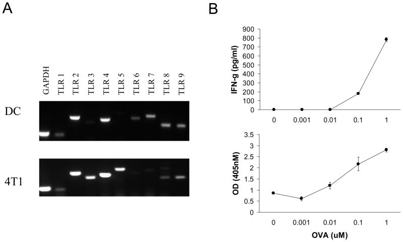
Fig. 1.
4T1 and CD11c+ DC express TLR2 and TLR4. A. RT-PCR was used to assess TLR1 to TLR9 expression by 4T1 and CD11c+ DC. GAPDH was used as a positive control. B. Functional activity of CD11c+ DC. OVA specific CD4+ T cells were cultured with OVA peptide pulsed DC to evaluate the antigen presenting capability of the DC. T cell cytokine secretion and proliferation were assayed at 48 hours. The data show the average and standard deviation of triplicate wells from a representative of each experiment. All data are representative of three separate experiments.

