
Figure 1.
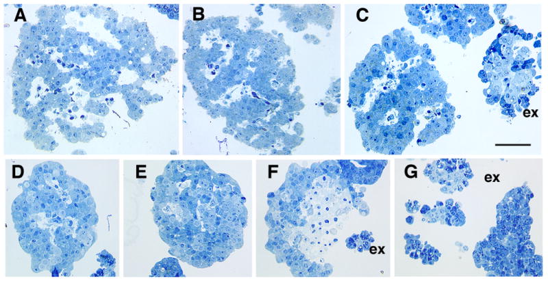
Identification of islet and non-islet tissue by light microscopy with plastic sections. Freshly isolated islet tissue is characterized by its cordlike pattern around vascular spaces (white areas) (A-C). These spaces partially collapse within 24 hr of culture at 37°C (D-G). Initially the vascular spaces of fresh human islets comprise about 14% of the islet volume (Pisania et al, submitted). Acinar cells (C, F,G) are distinguishable from the islets by their large zymogen granules (dark blue); the small terminal ducts (homogenous light blue) are seen surrounded by the acinar cells in these exocrine (ex) clumps (C,F,H). The exocrine clumps are initially compact (C) and do not show volume change with 24 hrs culture (G). Panel F shows necrosis of islet even after 24 hrs in culture. Toluidine blue stained one μm plastic sections of purified human islet preparations. Magnification bar = 50 μm.