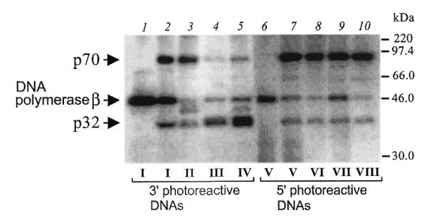
Figure 2.

p32 locates near the 3′-end of upstream oligonucleotide whereas p70 locates near the 5′-end of downstream oligonucleotide in DNA gaps. Primer elongation reactions using different template–primer structures (1 µM each) were carried out in the presence of NAB-4-dUTP for reagents I–IV or in the presence [α-32P]dCTP for reagents V–VIII (for conditions see Materials and Methods). After complete primer elongation 0.86 µM RPA was added to the reaction mixtures followed by UV irradiation. The numbers of the photoreagents (I–VIII, lanes 2–5, 7–10) used in each of the reaction mixtures are indicated at the bottom of the figure. Control reactions (lanes 1 and 6) did not contain RPA. The crosslinked protein–DNA complexes were separated by SDS–PAGE and visualized by autoradiography. The positions of crosslinked products and protein markers are indicated on the left and the right margins, respectively. Additional bands in lanes 3 and 5 appear to be products of modification of RPA proteolytic fragments since RPA preparation according to Coomassie staining shows minor bands that appeared during storage (data not shown).
