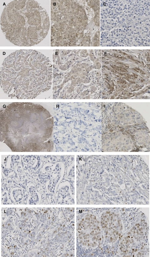
Figure 3.
Images of immunohistochemistry (IHC) for each antibody. (A–C) Breast cancer tissue stained with c-Src antibody (1 : 200, Cell Signalling). (A) An overview of a 0.6 mm core of the breast cancer tissue microarray, demonstrating no stromal staining, weak cytoplasmic, none and weak nuclear staining; magnification × 10. (B) Weak cytoplasmic, none and weak nuclear and weak membrane staining; magnification × 100. (C) Negative staining of stroma and tumour tissue; magnification × 100. (D–F) Breast cancer tissue stained with Lyn antibody (1 : 5, BD Biosciences). (D) An overview of a 0.6 mm core of the breast cancer tissue microarray, demonstrating no stromal staining, weak cytoplasmic, none and weak nuclear staining; magnification × 10. (E) Weak cytoplasmic, none and weak nuclear staining; magnification × 100. (F) No stromal staining, weak cytoplasmic, none, weak and moderate nuclear staining; magnification × 100. (G–I) Breast cancer tissue stained with Lck antibody (1 : 50, Cell Signalling). (G) Strong staining of tonsil with Lck (positive control); magnification × 2. (H) Negative staining of stroma and tumour tissue; magnification × 100. (I) Weak cytoplasmic and weak membrane staining; magnification × 100. (J–M) Ki67 staining of invasive breast cancer specimen (1 : 150, DAKO). (J) Negative staining of stroma and tumour tissue; magnification × 100. (K) Ki67 staining classified as weak staining; magnification × 100. (L) Ki67 staining classified as moderate staining; magnification × 100. (M) Ki67 staining classified as strong staining; magnification × 100.

