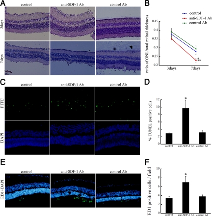
Figure 6.
Effect of SDF-1 inhibition on retinal ONL thickness. Anti-SDF-1 Ab or control Ab was intravitreally injected immediately after RD induction. The rats without intravitreal injections served as controls. A: No apparent morphological differences among the three groups, 3 days after RD. By day 7 after RD, ONL thickness in anti-SDF-1 Ab-injected eyes was significantly lower compared with normal controls or control Ab-injected eyes. Original magnifications, ×200. B: Graph summarizing effects of intravitreal injection on ONL thickness of rat retinas at 3 and 7 days after detachment. Results are the mean ± SEM. *P < 0.05; control Ab versus anti-SDF-1 Ab injection at day 7. **P < 0.01; control versus anti-SDF-1 Ab injection at day 7. C: TUNEL staining (green) and DAPI (blue) of detached rat retinas treated with anti-SDF-1 Ab or control Ab at day 3 after RD. D: Quantification of TUNEL-positive cells in ONL among three groups. Results are the mean ± SEM. *P < 0.05; control versus anti-SDF-1 antibody injection. E: Immunofluorescent staining for ED1 (green) and DAPI (blue) showing infiltrated cells into the subretinal space at day 7 after RD. Original magnifications, ×200. F: Quantification of the number of ED-1-positive macrophages among the three groups. Results are the mean ± SEM. *P < 0.01; control versus anti-SDF-1 antibody injection.

