Figure 4.
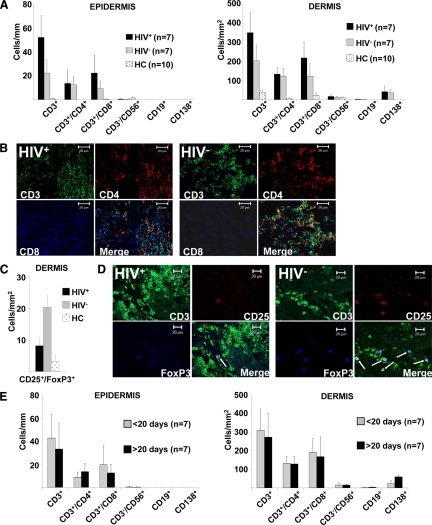
Alterations of the lymphocytic infiltrate in syphilitic lesions of HIV+ and HIV− patients. A: Detailed quantitative in situ analysis of lymphocytes within the epidermis and dermis of secondary syphilitic lesions and normal skin from healthy controls (HC). Single and double immunofluorescence staining was performed with the indicated markers from HIV+ and HIV− patients. Data are given as absolute numbers of positive cells ± SEM. B: Triple immunofluorescence labeling of syphilitic lesions from HIV+ and HIV− patients with anti-CD3 (FITC), anti-CD4 (TRITC), and anti-CD8 (APC) reveals an increase in CD8+ T-cells and total T-cells in biopsies from HIV-infected patients. Original magnification, ×400. C: Quantification of CD25+FoxP3+ T-cells by immunofluorescence double labeling. Data are given as absolute numbers of positive cells ± SEM. D: Triple immunofluorescence labeling of syphilitic lesions from HIV-infected and HIV− patients with anti-CD3 (FITC), anti-CD25 (TRITC), and anti-FoxP3 (Cy5) reveals decreased numbers of regulatory T-cells in HIV+ patients. Arrows denote triple positive cells. Original magnification, ×400. E: Quantification of cell subsets was performed by single and double immunofluorescence staining with the indicated markers. Samples were subdivided according their lesional age. Data are given as the absolute numbers of positive cells ± SEM.

