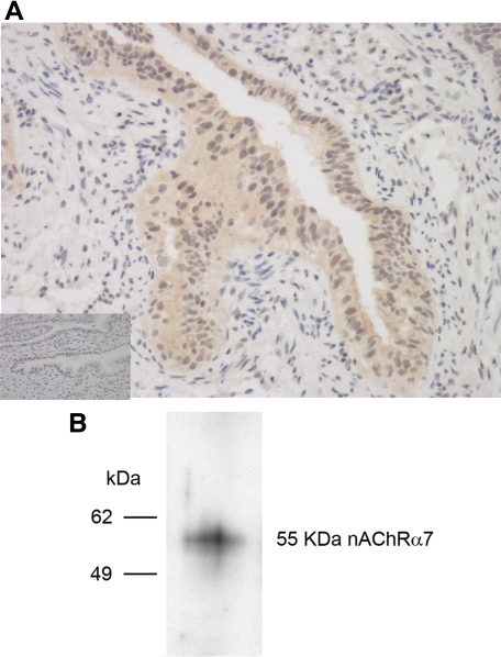
Figure 2.
Expression of nAChRα−7 in Fallopian tubes and OE-E6/E7 cells. A: Immunohistochemical staining of Fallopian tubes with rabbit polyclonal IgG antibody specific for nAChRα−7 (main image) and control rabbit IgG (inset) at ×100 magnification. B: Lysates of OE-E6/E7 cells resolved by SDS-PAGE, transferred to nitrocellulose, and blotted with a rabbit IgG antibody specific for nAChRα−7.

