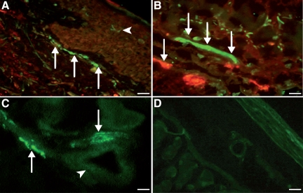
Figure 3.
Double-immunofluorescence staining for PACAP (polyclonal, green) and mast cell tryptase (monoclonal, red) in tissues from patients with urticaria (n = 6). A: Marked immunostaining for PACAP (green) in nerve fibers close to the dermal-epidermal border of human skin tissue (arrows). In the environment of epidermal nerve fibers, single PACAP-positive dendritic-like cells (Langerhans cells) of urticaria patients were detected (arrowhead, red; ×20; Scale bar = 40 μm). B: In the upper dermis, marked immunofluorescence staining for PACAP in nerve fibers (arrows) closely accompanied by tryptase-positive mast cells (red; ×40; Scale bar = 31.5 μm). C: PACAP-positive nerve fibers (arrows) close to dermal blood vessels (arrowhead, ×40; Scale bar = 31.5 μm). D: Negative control (pre-immune absorption control) showing absence of PACAP in nerve fibers and endothelial cells demonstrating specificity of immunostaining (×10; Scale bar = 45 μm).

