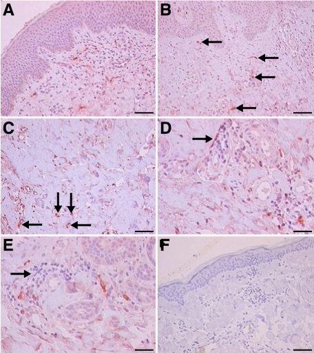
Figure 4.
Immunohistochemical detection of VPAC1R in urticaria human skin (n = 6). A: Overview shows intense dermal staining of VPAC1R in blood vessels and occasionally in leukocytes and weak staining for VPAC1R in keratinocytes and certain fibroblasts (×10; Scale bar = 45 μm). B: Higher magnification shows intense staining of endothelial cells, mast cells and lymphocytes for VPAC1R (arrows) (×20; Scale bar = 40 μm). C: Weaker staining for VPAC1R in the deeper dermis, small blood vessels, and leukocytes (arrows) as compared to superficial dermis (×40; Scale bar = 31.5 μm). D and E: Negative to weak staining for VPAC1R in dermal connective tissue, and occasionally staining of lymphocytes and mast cells (arrows, ×40; Scale bar = 31.5 μm). F: Control tissue (pre-immune absorption) demonstrates absence of VPAC1R in the skin, verifying specificity (×10; Scale bar = 45 μm). Experiments were performed as described in Materials and Methods.

