Figure 6.
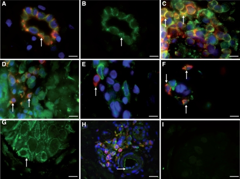
Double immunofluorescence of VPAC1R (green) in human skin sections of patients with atopic dermatitis (n = 6). A: Colocalization of VPAC1R and CD31+ in dermal vascular endothelial cells of atopic dermatitis patients (arrow). No staining of smooth muscle cells against VPAC1R was seen. B: Membrane staining for VPAC1R (green) in endothelial cells of the same postcapillary venule as Figure 6A (arrow). C: Colocalization of VPAC1R (green) in many, but not all, CD4+ T-helper cells (yellow, arrow) of dermal T-cell infiltrate (stained against CD4+, red). D: Immunofluorescence of VPAC1R (green) in the same infiltrate as (A), but now against CD8+ T-cells. Rare colocalization (yellow, arrow) of VPAC1R (green) in CD8+ cytotoxic T cells (red) of the infiltrate E: No colocalization of VPAC1R (green) in tissue macrophages within dermal infiltrate stained against CD68 (red, arrow). F: Weak colocalization (yellow) in a few mast cells stained for tryptase (red, arrow) and VPAC1R (green). G: Localization of VPAC1R in basal and suprabasal keratinocytes (green, arrow). H: Staining of endothelial cells (green) for VPAC1R (arrow). Note surrounding CD4+ T cells (red), colocalization in yellow. I: Control tissue (preimmune absorption) demonstrates absence of VPAC1R in the skin, verifying specificity. Experiments were performed as described in Materials and Methods. Magnification, ×100; Scale bars = 12 μm.

