Figure 1.
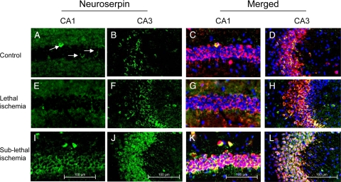
Sublethal ischemia induces neuroserpin expression in the hippocampal CA1 and CA3 layers. Representative micrographs from the CA1 and CA3 hippocampal layers in wild-type mice maintained under nonischemic conditions (controls, A–D) or 6 hours after either 20 minutes of BCCAO (lethal ischemia; E–H), or three episodes of BCCAO of 1 minute duration each with 5-minute intervals of reperfusion in between (sublethal ischemia, I–L) are shown. Green is neuroserpin, blue is DAPI, and red is the neuronal marker NeuN. Original magnification, ×20. Arrows indicate the presence of scattered neuroserpin-positive cells in the CA1 layer under normoxic conditions. Each observation was repeated five times.

