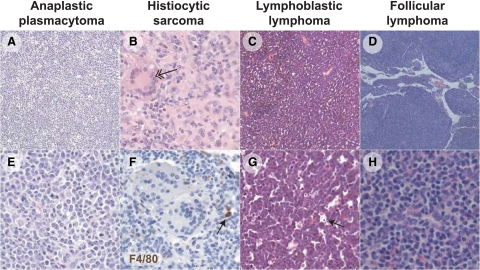
Figure 1.
Loss of Msh6 leads largely to B-cell lymphomas of diverse morphology. All panels were stained with H&E, except for F, which was stained by F4/80 (brown) and hematoxylin (blue). A: Tumor 1301 is an example of an anaplastic plasmacytoma that effaced the splenic follicular architecture with a uniform population of round cells (E) with ample pale basophilic cytoplasm with round nuclei (plasmacytoid) admixed with histiocytes. B: Tumor 624 is an example of a histiocytic sarcoma in which nuclei are large and oval, and large multinucleated forms are apparent (double arrow). F: The histiocytic-like neoplastic cells do not stain with the F4/80 antibody; a reactive macrophage in the micrograph (arrow) demonstrates effective immunohistochemistry with this antibody. C: Tumor 981 is an example of lymphoblastic lymphoma, with splenic architecture effaced by small dark round cells with uniform round stippled nuclei with one to two generally centrally placed nucleoli and scant cytoplasm (G). Throughout the lesions there were many tingible body macrophages carrying apoptotic bodies (arrow), endowing these tumors with a “starry sky” appearance. D: Tumor 1281 is an example of follicular lymphoma, in which neoplastic B cells have formed nodules composed of generally small cells with round-to-ovoid stippled nuclei, with numerous cells which are histiocytic in appearance (H). Original magnification: ×5 (D); ×10 (A and C); ×10 (B, E–H).

