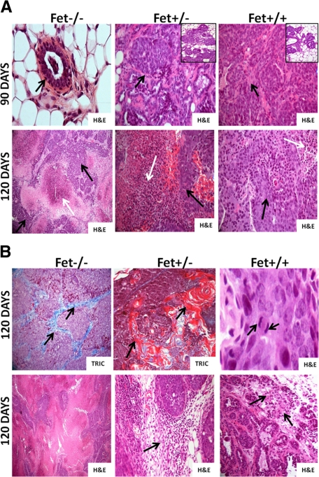
Figure 2.
Histopathology of mammary tissues of PyMT/Fet−/−, PyMT/Fet+/−, and PyMT/Fet+/+ at 90 and 120 days of age. A, top panel: Normal acinus (arrow) in a PyMT/Fet−/− mouse at 90 days of age (original magnification, ×40). The mammary tumor tissues of PyMT/Fet+/− and PyMT/Fet+/+ mice at 90 days of age clearly show tumor masses (arrows original magnification, ×20). There are also several hyperplastic acini and mammary intraepithelial neoplasm (insets). A, bottom panel: Tumors of PyMT/Fet−/− (original magnification, ×10), PyMT/Fet+/−, and PyMT/Fet+/+ (original magnification, ×20) mice show expansile tumor masses and extensive to moderate areas of necrosis (white arrows) interspersed with islands of neoplastic cells (black arrows). B, top panel: Mammary tumors show accumulation of collagen fibers particularly in tumors in PyMT/Fet−/− mice (stained blue with trichrome [TRIC], arrows). Tumor tissues of PyMT/Fet−/− and PyMT/Fet+/− also show squamous metaplasia and parakeratosis with concentric lamellar keratin consistent with keratin pearls (stained red with trichome; original magnification, ×20). Mitotic figures (arrows) for PyMT/Fet+/+ tumors of 3 to 5 per high-power field are shown (original magnification, ×40). B, bottom panel: tumor tissues in PyMT/Fet−/− mice show a medullary pattern (original magnification, ×10). This panel also shows mild to moderate mononuclear cell infiltration (arrows) in the tumor tissues of PyMT/Fet+/− and PyMT/Fet+/+ mice (original magnification, ×20).

