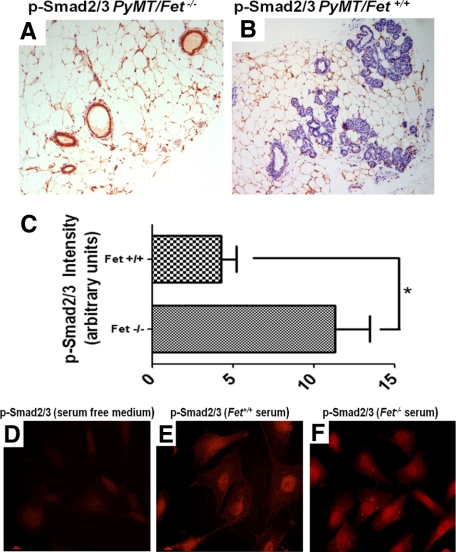
Figure 4.
TGF-β signaling determined by staining for P-Smad2/3 in mammary tissues of mice and human cells. A and B: Intensity of P-Smad2/3 staining in the mammary tissues of 60-day-old PyMT/Fet−/− (A) and PyMT/Fet+/+(B) mice (original magnification, ×10). C: Intensity of P-Smad2/3 staining represented as arbitrary units. *P = 0.008 D–F: TGF-β signaling in BT-549 breast tumor cells incubated in the absence of serum (D) and in the presence of serum from PyMT/Fet+/+ (E) and PyMT/Fet−/− (F) animals.

