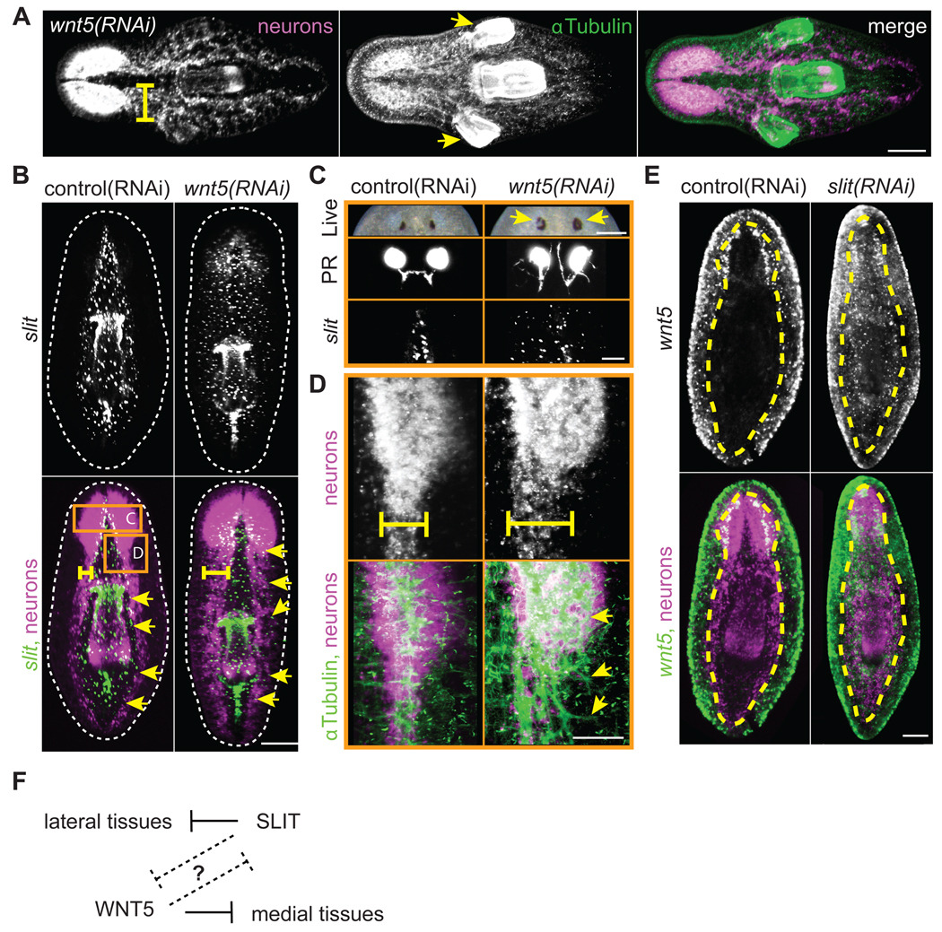
Fig. 6.
WNT5 and SLIT reciprocally pattern the mediolateral axis. (A–E) Neurons; PC2 riboprobe (magenta in merges) (A) wnt5(RNAi) trunk fragments exhibited laterally expanded cephalic ganglia, defasciculated axon tracts (yellow bar), and ectopic lateral pharynges (yellow arrows) by 15 dpa. Brain, axons, and pharynx; α-Tubulin antibody (green in merge). (B) Lateral expansion of the midline in trunk fragments 15 dpa. Midline; slit riboprobe (green in merge). Yellow bar; width of VNCs. Yellow arrows; lateral extent of slit expression. Dotted line; periphery of animal. Note that slit expression was bounded by the VNCs in control animals. (C–D) Higher magnification of regions boxed in panel B. Regenerating wnt5(RNAi) trunks at 15 dpa display (C) ectopic pigment (yellow arrows) around the photoreceptor (PR) pigment cup (live image is 22 dpa) and aberrant PR axon projections. PR axons; VC-1 antibody. Midline; slit riboprobe. (D) Axons; α-Tubulin antibody (green in merge). Note that wnt5(RNAi) axon tracts were defasciculated and disorganized (yellow arrows). (E) slit(RNAi) caused ectopic expression of wnt5 (green in merge) across the midline. Yellow dashed line; outer edge of the ventral nerve cords. (F) Model. WNT5 and SLIT act reciprocally, either directly or indirectly, to properly pattern the mediolateral axis. Scale bars; (A–B, E) 200µm, (C,D) 100µm.

