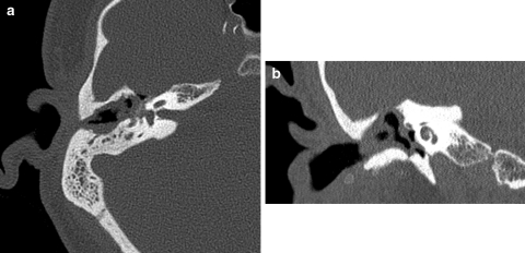
Fig. 1.
Axial (a) and coronal (b) CT scan of the temporal bone demonstrating soft tissue density with extension from the right external auditory canal to the right epitympanum, mesotympanum, and hypotympanum with dehiscence of the overlying right tegmen tympani and absence of the right ossicular chain

